Understanding Ventriculomegaly in the Fetal Brain
This blog is designed to help you understand what ventriculomegaly is, why it can occur during pregnancy, what tests you may be offered, and what it may mean for your baby. We understand that receiving this information can be worrying, and we hope this guide helps answer your questions.
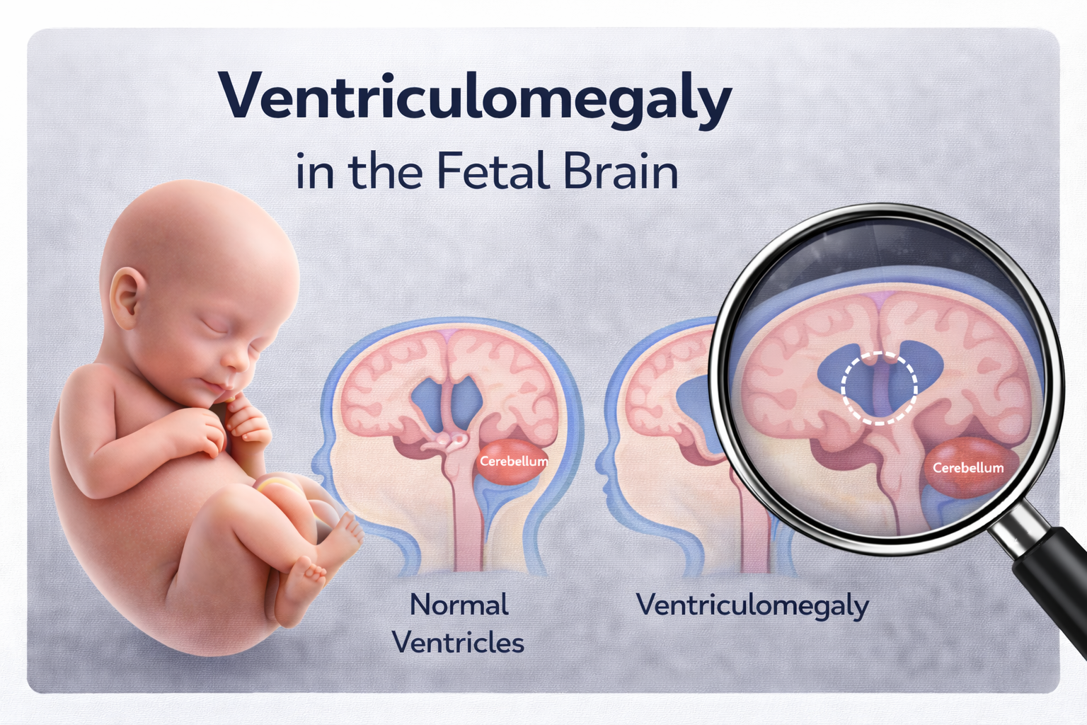
What Is Ventriculomegaly?
During routine pregnancy scans, the baby’s brain is carefully examined. Inside the brain are small spaces called ventricles, which are filled with cerebrospinal fluid (CSF). This fluid cushions the brain and spine and helps them develop normally.
Two of these ventricles—one on each side of the brain—can be clearly seen and measured on ultrasound to check their size:
- Up to 10 mm: normal
- 10–15 mm: ventriculomegaly
- Above 15 mm: hydrocephaly
Why Does Ventriculomegaly Happen?
There are several reasons why the ventricles may appear wider than usual:
- The fluid inside them may not flow as smoothly as it should.
- An infection or a difference in the development of the brain or spine can affect fluid movement.
- In some cases, changes in the baby’s chromosomes (which carry genetic information) may be involved.
- Very rarely, bleeding into the baby’s brain can cause the ventricles to enlarge.
It is important to remember that many babies with ventriculomegaly are otherwise healthy,
and often no clear cause is found. When no cause is identified, the condition is called isolated ventriculomegaly.
What Tests Might Be Required?
If your baby’s ventricles measure above 10 mm, your healthcare specialist may suggest further tests to understand whether there is an underlying reason for the enlargement:
1. Detailed ultrasound scan
A specialist will look carefully at your baby’s brain and the rest of the body, especially the heart, to check for any associated findings.
2. Amniocentesis
A small sample of the fluid around your baby is taken. This can be tested for:
- Chromosomal conditions
- Single-gene differences
- Certain infections
3. Infection screening
A blood test may be offered to check whether infections such as CMV or toxoplasmosis could have contributed.
4. Fetal MRI
Later in pregnancy, an MRI scan may be offered to look at the baby’s brain in even greater detail.
Is Genetic Testing Required in Fetal Ventriculomegaly?
Genetic testing is an important part of evaluating fetal ventriculomegaly. It helps determine whether the enlarged ventricles are related to chromosomal or genetic conditions, such as Down syndrome, trisomy 13 or 18, microdeletions or microduplications, or certain single-gene disorders (especially in male fetuses, including some X-linked conditions).
When all genetic tests are normal and no other abnormalities are found, the condition is considered isolated ventriculomegaly, and the outlook is generally reassuring— particularly when the enlargement is mild.
A range of genetic tests may be used, including FISH, Q-f PCR, Karyotyping, Microarray analysis, and whole-exome sequencing. The final decision about which tests are most suitable will be made after evaluating multiple factors relevant to your case.
Types of Genetic Tests Suggested
• FISH: Quick test for major chromosomal abnormalities.
• QF-PCR: Rapid test for common chromosome conditions (e.g., Down, Edwards, Patau syndromes).
• Karyotyping: Looks at the full set of chromosomes for large changes.
• Chromosomal Microarray: Detects small missing or extra pieces of DNA (microdeletions/duplications).
• Whole Exome Sequencing (WES): Analyses many genes to detect more specific genetic conditions.
The choice of test depends on several factors, including the degree of ventriculomegaly, whether other abnormalities are seen, and specialist recommendations.
How is the fetus monitored?
You will usually be offered follow-up ultrasound scans to check whether the ventricles:
• Stay the same size
• Become smaller
• Increase in size
Many parents find this ongoing monitoring helpful and reassuring.
What Does This Mean for Baby After Birth?
The outlook depends largely on whether a cause is found. If the ventriculomegaly is isolated and mild:
• The outcome is often good, especially if the ventricular size remains stable throughout pregnancy.
• Babies with mild, isolated ventriculomegaly frequently develop normally.
However, proper post-birth evaluation with the appropriate specialists and regular follow-up are strongly recommended to ensure your baby receives the best possible care.
The chance of ventriculomegaly recurring in a future pregnancy depends
on whether a genetic cause was identified. Your healthcare team or a genetic counsellor can discuss this with you in more detail.
- Choroid Plexus Cyst
- Who is at Risk?
- Down Syndrome
- Thickened Nuchal Fold in Fetus
- Absent or Small Nasal Bone
- Echogenic intracardiac focus (EIF) in the fetal heart
- Cerebellar hypoplasia (CH) in Fetus
- Agenesis of Septum Pellucidum in Fetal Brain
- Ventriculomegaly in the Fetal Brain
- Mega Cisterna Magna – MCM
Committed to precise diagnostics, empathetic guidance, and advanced fetal care — supporting mothers and babies at every step of their journey.
Quick Links
Contact Information