Understanding Agenesis of Septum Pellucidum in Fetal Brain
This blog explains what agenesis of the septum pellucidum (ASP) is, the tests you may need, and what it could mean for your baby .
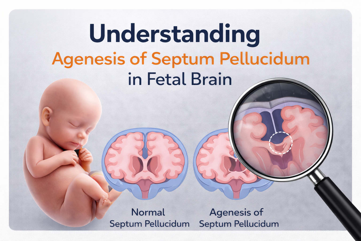
What is Agenesis of the Septum Pellucidum?
The septum pellucidum is a thin wall in the middle of the brain that separates two fluid-filled spaces called the lateral ventricles.
Agenesis of the septum pellucidum (ASP) means that this wall is missing or not seen on an ultrasound. Normally, the septum pellucidum:
• Attaches to the corpus callosum (connects the left and right sides of the brain) at the top.
• Connects to the fornix (a bundle of nerves) at the bottom. ASP can sometimes be associated with changes in:
• Nerves to the eyes
• The pituitary gland, which produces hormones
How does it happen?
Most cases occur by chance, and the exact cause is often unknown. Possible causes include:
• Genetic changes, due to Chromosomal and Single defects
• Infections or chemical exposures during pregnancy
• Other brain anomalies that prevent the septum pellucidum from forming
Tests You May Be Required
If ASP is suspected, your doctor may recommend:
1. Detailed fetal brain ultrasound – Neurosonogram
2. Fetal MRI (magnetic resonance imaging) – provides different views of the brain and surrounding structures and is safe during pregnancy
3. Fetal Genetic testing
Why Genetic Testing is Performed
An abnormal or absent CSP sometimes indicate an underlying genetic syndrome. Conditions linked to CSP abnormalities include agenesis of the corpus callosum, holoprosencephaly (HPE), trisomy 13, 18, and 21, 22q11.2 deletion syndrome, and Cri du chat syndrome. Identifying a genetic cause can help determine
the likely prognosis and guide management decisions during pregnancy and after birth. Even when the CSP appears isolated, a genetic investigation is often recommended to rule out subtler chromosomal or genetic abnormalities.
What Type of Genetic Testing is required?
Genetic testing typically begins with a diagnostic procedure to obtain fetal cells.
This can be done through amniocentesis, where a sample of amniotic fluid is taken.
Once fetal cells are obtained, several analyses may be performed
• Karyotype analysis: Detects large-scale chromosomal abnormalities.
• Fluorescence in situ hybridization (FISH): Identifies specific chromosomal changes, such as deletions in the 22q11.2 region.
• Chromosomal microarray analysis (CMA): Provides a more detailed assessment, detecting smaller chromosomal changes, that might be missed by a standard karyotype.
• WES: whole exome sequencing has been shown to increase the diagnostic yield (the rate at which a specific genetic cause is found) in fetuses with
central nervous system (CNS) abnormalities when standard tests like karyotyping and chromosomal microarray (CMA) are negative.
During Pregnancy
• No special changes to routine pregnancy care are usually needed
• Ultrasounds scans may be repeated later in pregnancy to check for any changes in the brain
After Birth: What It Means for Your Baby
The outcome depends on whether other brain abnormalities are present:
• If other severe anomalies exist: The baby may have significant health issues.
• About 25% of cases: Problems with the eyes or pituitary gland can occur, possibly affecting vision or hormone production. Most children will still have normal intelligence.
• If ASP is isolated (no other brain changes): The baby’s outcome can be better .
Risk of Recurrence
If there is no genetic cause, the risk of it happening again in future pregnancies is very low
- Choroid Plexus Cyst
- Who is at Risk?
- Down Syndrome
- Thickened Nuchal Fold in Fetus
- Absent or Small Nasal Bone
- Echogenic intracardiac focus (EIF) in the fetal heart
- Cerebellar hypoplasia (CH) in Fetus
- Agenesis of Septum Pellucidum in Fetal Brain
- Ventriculomegaly in the Fetal Brain
- Mega Cisterna Magna – MCM
Committed to precise diagnostics, empathetic guidance, and advanced fetal care — supporting mothers and babies at every step of their journey.
Quick Links
Contact Information